 ARIA -
Ratings explained ARIA -
Ratings explained |
Appropriateness
of Referral and Investigation in Angina (ARIA)
1.
Introduction
Two panels consisting of 5 General Practitioners, 5 Cardiologists and 2
cardio-thoracic surgeons met in Summer 2003 to rate 1128 indications
(combinations of clinical and patient characteristics) for
appropriateness of Exercise testing and 3071 indications for coronary
angiography.
Each indication was rated on a
scale from 1 to 9, 1-3 denoting inappropriate, 4-6 equivocal and 7-9
appropriate.
The judgements of the panellists
were based on the balance of clinical benefit : harm for each
individual patient constellation based on the best available research
evidence and their own expertise. It was assumed that cost criteria did
not apply, there were no waiting lists and every clinician had
immediate open access for each investigation. These assumptions were
made because the ratings should capture the clinical necessity of an
investigation that is applicable universally and over time. Factors
such as access restrictions or waiting lists are considerations that a
clinician has to take in the context of clinical appropriateness.
Sometimes this will mean that investigations cannot be carried out even
though the patient remains clinically appropriate.
For the same reason the patient
was assumed to be free of major comorbidity, absolute indications,
absolute contra-indications or preference. The presence of any of these
factors may justifiably override any appropriateness for exercise ECG
and coronary angiography. A patient who refuses an angiogram will not
receive it despite the potential benefit of angiography.
2. Factors thought to influence appropriateness of testing
An indication is defined by several clinical descriptors such that each
patient satisfying one indication is equally appropriate (or
inappropriate) for a given management step.
Six broad clinical presentations
Based on clinical practice, recommendations from guidelines and
published research, the ARIA indications are grouped into six broad
clinical presentations.
1
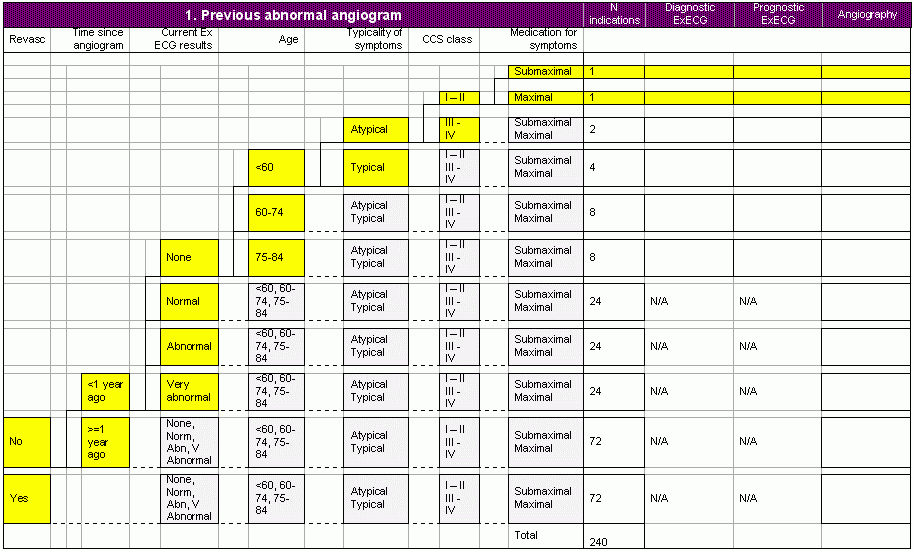
Previous abnormal coronary angiogram.
This is defined as the presence of at least one vessel with at least a
50% stenosis.
2
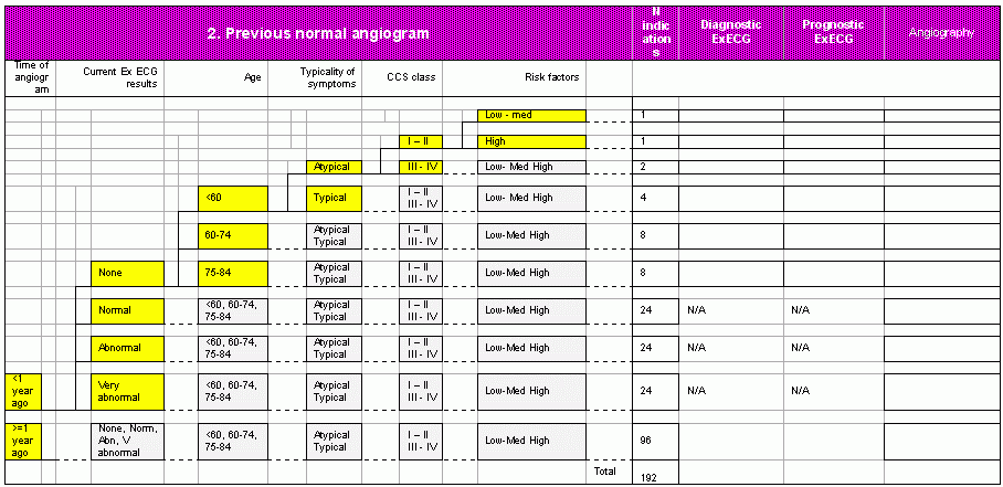
Previous normal coronary angiogram.
This is defined as absence of any luminal narrowing.
3.
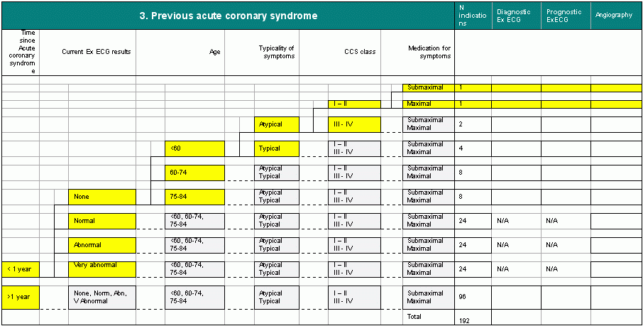
Previous acute coronary syndrome (acute myocardial infarction or
unstable angina)
Defined on the basis of typical history and electrocardiographic
changes or biochemical changes. Thus the acute coronary syndrome is
“definite,” not based on history alone. Thus in these patients the
diagnosis of underlying coronary disease is in little doubt.
4.
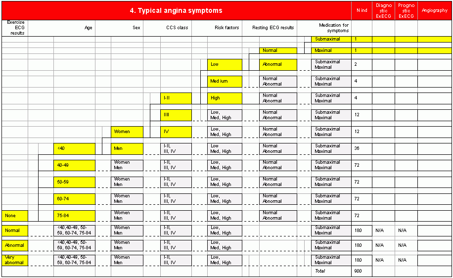
Typical angina symptoms
Pain or
discomfort accompanied by all 3 of:
Site:
Retrosternal, arms, chest
Precipitating factors: Exercise/cold/emotion
Relieving factors: Rest or GTN
5.
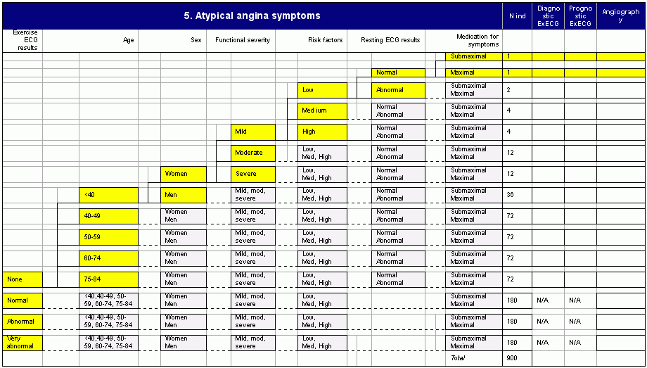
Atypical anginal symptoms
Pain or discomfort accompanied by any 2 out of 3
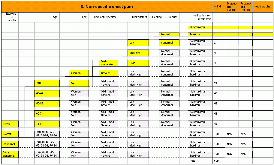
6.
Non-specific chest pain
Pain or discomfort accompanied by any 1 out of 3
N.B. This widely used definition, does not include time to relief, or
nature of chest pain.
For
presentations 3-6, no patient has previously undergone angiography.
For presentations 4-6 , no patient
has a history of acute myocardial infarction or unstable angina.
Ten specific factors defining indications
Within each of
the 6 broad clinical presentations, specific indications are defined
using some or all of the following clinical descriptors.
1. Previous revascularisation. Either PCI or CABG, assumed
to be complete revascularisation.
2. Time since angiography. Self explanatory
3. Time since acute coronary syndrome. Self explanatory.
4. Age. A maximum of 5 levels is considered (<40, 40-49,
50-59, 60-74, 75-84)
5. Sex
6. Severity of symptoms. Canadian Cardiovascular Society
(CCS) scale is used for defining angina severity71. The CCS class is
defined according to the box. In practice panellists may choose to
consider a more general classification of mild (CCS class I-II),
moderate (III) or severe (IV). The severity of symptoms is current,
once the patient is on anti-anginal medication.
| CCS I |
“Ordinary
physical activity does not cause…angina,” such as walking and climbing
stairs. Angina with strenuous or rapid or prolonged exertion at work or
recreation. |
| CCS
II |
“Slight
limitations of ordinary activity.” Walking or climbing stairs rapidly,
walking uphill, walking of stair climbing after meals, or in cold, or
in wind, or under emotional stress, or only during the few hours after
awakening. Walking more than 2 blocks on the level and climbing more
than one flight of ordinary stairs at a normal pace and in normal
conditions. |
| CCS
III |
“Marked
limitation of ordinary physical activity.” Walking one or two blocks on
the level and climbing one flight of stairs in normal conditions and at
normal pace. |
| CCS
IV |
“Inability
to carry on any physical activity without discomfort – anginal syndrome
may be present at rest.” |
Functional
severity of non-specific chest pain
For patients with non-specific chest pain it makes no sense to describe
a CCS class. However such patients do differ in their functional
limitations from their pain. For the purposes of rating this is defined
at two levels as mild – moderate or severe. This mirrors clinical
judgement in which some patients present with non-specific chest pain
have severe functional impairment, or represent an important clinical
problem because of repeated admissions.
7. Risk
factors
Those factors which add incremental diagnostic or prognostic
information to knowledge of age, sex and clinical presentation are
termed “risk factors”.
As the
evidence commentary shows, there are no scores validated in primary
care which demonstrate how simple risk factors combine to contribute
incremental diagnostic or prognostic value. The Framingham risk scores
are not relevant because people with angina at baseline were excluded
from the cohort.
Risk is
defined simply, and partly implicitly, as low, medium or high based on
smoking, hypertension, lipids, family history, obesity, south Asian
ethnicity, diabetes and arterial disease in non-coronary circulations.
Note age and sex are considered separately. Clinicians vary in which
combination of risk factors they denote as “high risk” but for purposes
of standardising the panel discussion the following definitions should
be used:
Low risk “0”
risk factors
Medium risk 1-2 risk factors
High risk 3 or more risk factors or
diabetes or
proven arterial disease in cerebral or peripheral circulations
8. Resting ECG abnormality
Is defined by the presence of pathological Q waves, or regional ST
depression, or regional T wave inversion. These resting ECG
abnormalities are considered to be ischaemic in origin.
9.
Exercise ECG
All exercise ECG results are current i.e. the test was carried out at
the time of the current presentation with symptoms. The following
definitions have been used by RAND,72 and the ACRE study13 in previous
expert panels rating the appropriateness of coronary angiography.
a. None
when no current exercise ECG results are available
b. Normal
The absence of a very abnormal or abnormal test (see below) and any one
of the following achieved:
· at least 85 percent of the predicted maximum heart rate
· heart rate/blood pressure product (heart rate x systolic
arterial pressure + 100) of 250 greater
· 10 METS
· completed stage IV
c. Abnormal
After the first 3 minutes of the test the patient develops
(i) 1 mm or more of horizontal or downsloping ST segment depression
that is present 80 msec after the J-point or
(ii) typical angina
d. Very abnormal
(i) During the first 3 minutes of the test (or onset at heart rate less
than 120 beats/minute, off beta-blockers, or less than 6.5 METS) the
patient develops
§ 1 mm or more of horizontal or downsloping ST segment
depression that is present 80 msec after the J-point or
§ typical angina or
(ii) Based on blood pressure criteria, either of the following
§ exercise test stopped because of a fall in blood pressure or
§ test read as abnormal and at least two consecutive 10mm blood
pressure systolic drops from the baseline (defined as the lower of
standing or hyperventilation systolic blood pressure) which occurs
during the exercise phase of the test.or
(iii) More than 2 mm of horizontal or downsloping ST depression at
any time or
(iv) Persistence of ST depression greater than 6 minutes post exercise
N.B. An
indeterminate test is not separately considered in the indications, on
the grounds that it is equivalent to not done.
10.
Medication for symptoms is defined as either sub-maximal or maximal.
Sub-maximal is defined as only one agent, or two agents at sub-maximal
dose. Maximal is defined as two or more agents at maximal dose, such
that the physician has no further therapeutic option with drugs alone
in the control of symptoms.
The
structure of the indications is outlined on the following pages.
3.
Ratings process
The RAND – Delphi method of rating the appropriateness of indications
for referral and investigation of involve two rounds the first, in
private, and the second where the panel members meet. All 22 panellists
received software, ratings protocol and a review of the best available
evidence and rated indications in private.
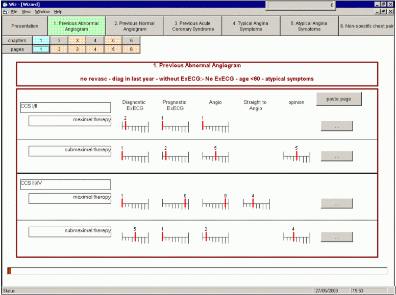
The software for rating has been
developed by the ARIA investigators. The layout of the software is
shown in Figure 1.
Figure 1 ARIA Wizard. Examples of
indications and ratings: no previous ECG
After returning the ratings the scores from each panel member were
collated for each indication and a personalised report on paper was
generated. This showed the panellists own rating, the median of the
whole panel, the range of the whole panel or whether the panel on the
whole agreed or disagreed on an indication. Disagreement for an
indication-decision pair is defined when:
Median of the panel =
appropriate, but at least 2 panellists rate it inappropriate
or
Median of the panel = inappropriate, but at least 2 panellists rate it
appropriate.
Panellists also received an
agenda which highlighted the main areas for discussion at the two day
panel meeting. The discussion focussed on important areas of
disagreement and important areas of agreement, if the panels were
grossly at odds with existing guidelines. The discussion aimed to
achieve consensus and were asked to rerate indications if they were
persuaded by the discussion. This process also helped eliminate any
discrepancies due to misinterpretation or misunderstanding.
These ratings present the
first appropriateness ratings for investigation in patients with angina
by two independent panels.
This page last modified
21 October, 2005
by Web
Administrator
|
