Tiling Images
Using a tiling microscope stage and software it is possible to obtain
images of large section areas whilst retaining a very high resolution. This
enables images to show both the gross cerebellar architecture and cellular
resolution required for detailed anatomical analysis.
Since these high resolution images are extremely memory intensive,
especially for download on the Internet, the examples shown are resampled at
low resolution.
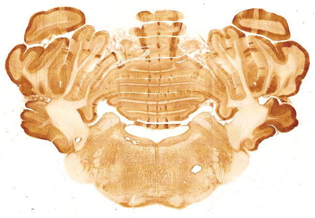
The above image is made up of 48 images. These
have been automatically tiled together so that the joins are near invisible.
The software automatically detects corresponding structures and glues the
pieces together on the fly as the stage moves from one field to another.