Regional brain shrinkage in MS predicts disability
16 February 2018
A UCL-led research team has identified the pattern of brain tissue loss in multiple sclerosis, enabling improved prediction of disability progression.

The study, published in Annals of Neurology, was one of the largest brain imaging studies ever conducted investigating multiple sclerosis (MS).
"It's well known that brain atrophy occurs in people with MS and varies by region, but we typically only measure the shrinkage of the whole brain. By looking at brain tissue loss in a more detailed fashion in different MS subtypes, we found it's possible to predict disability progression in advance. This has many implications in clinical trials that use brain atrophy to investigate the effect of neuroprotective drugs," said the study's lead author, Dr Arman Eshaghi (UCL Institute of Neurology).
The researchers collected data retrospectively from seven research centres in the UK, Italy, Spain, Austria and the Netherlands involved in the MAGNIMS consortium, pulling together MRI scans and disability measures from 1,417 study participants (1,214 with MS), with an average follow-up of 2.41 years.
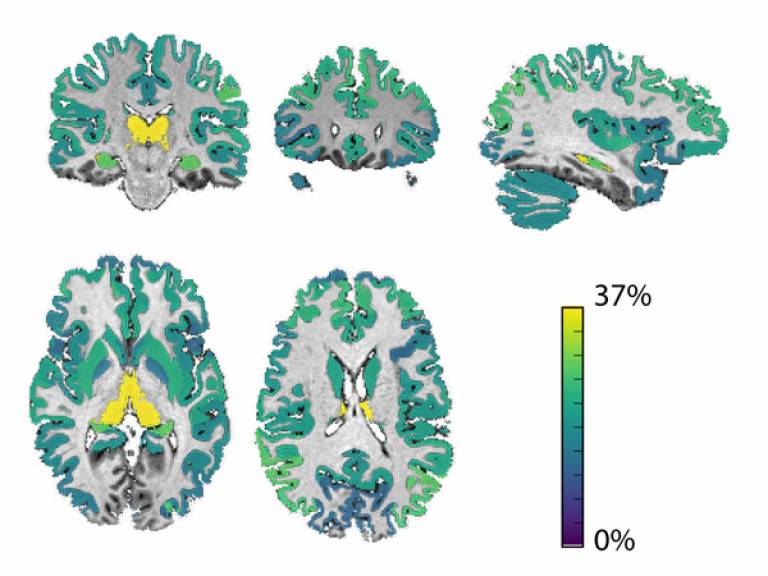
The research team found that loss of volume of the deep grey matter (which is deep in the brain, or the subcortex) was faster than loss of other brain regions. They also found a correlation between deep grey matter volume loss and disability accumulation. The strongest relationship was found in the thalamus, the largest component of deep grey matter, which is involved in relaying sensory and motor signals.
The study identified multiple spatial patterns of grey matter atrophy, including some that are dependent on type of MS, which is a highly heterogeneous disease. They found evidence suggesting that regions that lie more deeply in the brain and have many connections may be more vulnerable to atrophy than other regions.
This study shows the pattern of ongoing brain shrinkage in different MS subtypes for the first time with far reaching implications for development of future treatments in MS.
"Our methods could be used right away for Phase II clinical trials, offering up a higher sensitivity than existing measures to investigate whether a new drug could work," said Dr Arman Eshaghi (UCL Institute of Neurology).
The researchers caution that the method does not help predict disability progression in individual patients; rather, it's useful at the population level, providing a valuable measure for use in clinical trials of putative treatments that protect nerve cells.
"We have demonstrated that deep grey matter atrophy is linked to disability progression in multiple sclerosis by using advanced imaging analysis techniques and collecting a large number of MRI scans, because of the effort of many European researchers," said Professor Olga Ciccarelli (UCL Institute of Neurology), the senior author of the study.
The study was conducted by researchers at the UCL Institute of Neurology, the UCL Centre for Medical Imaging Computing, the National Institute for Health Research University College London Hospitals Biomedical Research Centre, the London School of Hygiene & Tropical Medicine, the University of Siena, S Camillo Forlanini Hospital, the University of Rome Sapienza, Vita-Salute San Raffaele University, Universitat Autonoma de Barcelona, VU University Medical Centre, Medical University of Graz, University of Pavia, and the C.Mondino National Neurological Institute as part of the MAGNIMS consortium.
The research team received funding from MAGNIMS, Multiple Sclerosis International Federation, the National Institute for Health Research University College London Hospitals Biomedical Research Centre, ECTRIMS, the EPSRC, the European Union's Horizon 2020 research and innovation programme and the Michael J. Fox Foundation for Parkinson's Research.
Links
- Research paper in Annals of Neurology
- Professor Olga Ciccarelli's academic profile
- UCL Institute of Neurology
- UCL Centre for Medical Imaging Computing
Image
- Brain areas with the strongest link between grey matter loss and risk of disability accumulation, with the thalamus shown in yellow. Credit: A Eshaghi et al
Media contact
Chris Lane
Tel: +44 (0)20 7679 9222
Email: chris.lane [at] ucl.ac.uk
 Close
Close

