Regenerative medicine success for muscles
31 March 2011
Links:
 ucl.ac.uk/ich/" target="_self">UCL Institute of Child Health
UCL Centre for Stem Cells and Regenerative Medicine
An
innovative strategy for regenerating skeletal muscle tissue using cells from
the recipient's own body is outlined in UCL research published today.
The paper, authored by Dr Paulo de Coppi (UCL Institute of Child Health and surgeon at Great Ormond Street Hospital) and colleagues, shows that damaged muscle tissues treated with satellite cells in a special degradable hydrogel showed satisfactory regeneration and muscle activity. Muscle activity in repaired muscle in a mouse model was comparable with untreated muscles. This is the first time muscle function has been proved by physiological tests.
The research is published in the Journal of the Federation of American Societies for Experimental Biology and represents an impressive development in the growing field of regenerative medicine.
Satellite cells (SCs), freshly isolated or transplanted within their niche, are presently considered the best source for muscle regeneration. They are located around existing muscles. Hence, a patient's own cells can be used, from a muscle biopsy.
A key issue for regeneration is how cells grow as a structure, as they usually require some form of framework. A hard framework would impede muscle growth and muscle cell penetration. The hydrogel, by contrast, provides a supportive structural skeleton but degrades quickly as muscle tissue returns and the support becomes unnecessary. The gel is initially liquid, hardens in place under UV light, and is easily penetrated by muscle cells.
"We focused on a simple, robust, and reproducible technique that could be easily adapted to clinical requirements" said Dr De Coppi.
"This is using the patients own cells, without any lengthy culturing process, which means we could take a biopsy, produce the cells in a couple of hours, and implant them where needed - it can be done in theatre as one process. Using the patient's own cells eliminates any tissue rejection."
"Nor are there ethical debates as for embryonic stem cells, or culturing and availability issues, such as amniotic stem cells."
The lab model has the potential to be translated into
significant clinical benefit for babies and children born with defective
organs, or caused by injury or pathological conditions, that currently require complicated and potentially devastating reconstructive
surgery.
Professor Andrew Copp, Director of the UCL Institute of Child Health, said: "This is a very exciting research finding that may significantly advance our ability to repair muscle damage or defects in future. It is a great example of translational research in action, from the laboratory to a near-clinical application."
The focus for initial clinical research in humans will be relatively small muscles at first, like deformities in the face and palate, or in the hand. It will be technically more demanding to grow larger muscles with more structure, which would require their own nerves and blood supply.
"We want to move to safe and effective human trials" said Dr de Coppi, "but of course we are not there yet. Furthermore scaling up from small muscles to larger structures will undoubtedly be challenging."
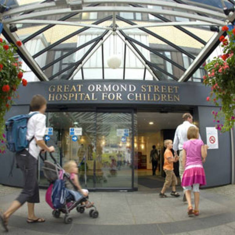
Image: Great Ormond Street Hospital for Children.
UCL context
The UCL Institute of Child Health, in partnership with Great Ormond Street Hospital, is the largest centre in Europe devoted to clinical and basic research and postgraduate teaching in children's health.
Great Ormond Street Hospital, along with four other of Britain's world-renowned medical research centres and hospitals, is part of UCL Partners, a collaboration of world-class researchers and clinicians to create Europe's strongest academic health science partnership, focused on preventing and treating major diseases that affect the populations in London, the UK and worldwide.
Related news
£28
million boost to UCL-led birth cohort study
New
research: infant nutrition and obesity
 Close
Close

