
What is Biomechanics and How Relevant is It to Healthcare?
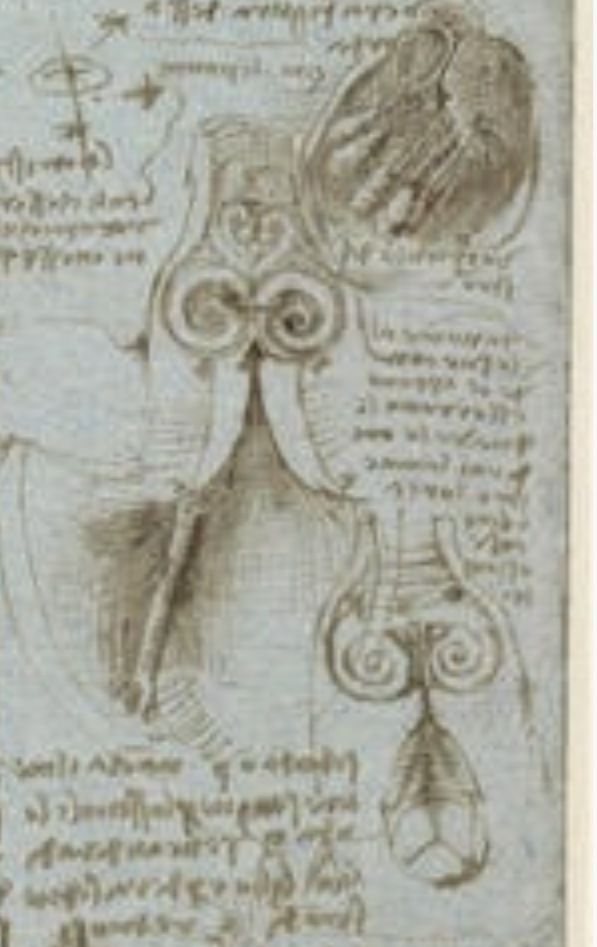 Biomechanics is one branch of study aimed at understanding the functionalities of biological systems using the knowledge of mechanics. This includes flow, structure, mass transport, etc. across scales from cellular (or even sub-celllular, =< 1 micron), organ (~1cm) and whole body (~1m) levels. Biomechanics has a rather long history. For example, Leonardo da Vinci in 16th century applied knowledge of fluid and solid mechanics to understand the functionality of human body. This can be seeen in his drawing of the flow patterns through human heart valve (figure [2]), which has been confirmed by many studies in vivo [3] and in vitro [4]. UK academia has made a number of landmark contributions in the field of biomechanics, such as the detailed description of blood flow (systemic) circulation in our body by Harvey [5]. Caro's low shear stress theory in the process of atherosclerosis [6] is a more recent example, which has driven many studies on atherosclerosis in these days, including ours (please see Coronary Artery Disease page for details). Obviously, the functionalities of living organisms are not only made of mechanics, hence it is vital to understand the interaction between the biomechanical and biological systems. In cardiovascular systems, which is our primary interest, mechanical factors often give a cue to biochemical processes that is key parts of its mechanism in health and disease. Many of our research projects are based on this, aimed at deriving biomechanical risk factors/indicators, such as wall shear stress acting on the vascular endothelium, to predict disease progression and clinical events.
Why Computational Biomechanics?
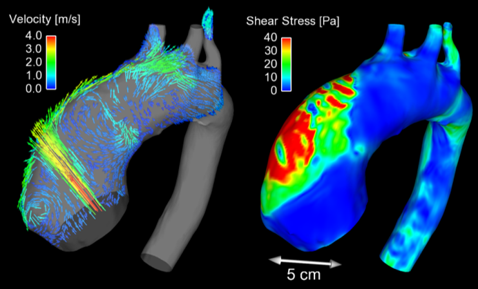 Some of the biomechanical variables, e.g. blood pressure, can be measured directly but many others are difficult to be measured. Haemodynamic wall shear stress acting on the vascular endothelium is a typical example of this. Even in case direct measurement is possible, it may not be easy if it is to be done in blood vessels deep in the human body. Typical examples include blood pressure measurement in coronary arteries, which requires a pressure-sensing cathether probe inserted in the coronary arteries, typically through an artery in groin. Computational Biomechanics can be used to calculate such biomechanical metrics based on patient's anatomical and other physiological data, in combination with fluid, solid and/or other multiphysics simulations. Computational derivations of biomechanical metrics require much less of invasive sensors/measurements, and could work as an alternative non-invasive "measurement" method of human pathophysiologcal states. Recent success of computationally-derived coronary Fractional Flow Reserve for the severity assessment of coronary atherosclerotic narrowing is an excellent example of this [7-10]. At the same time, model assumptions are inevitable for computer simulations and it is vital to know the principles and details of such methodologies, so that we can make the best out of the simulations. Overall, Computational Biomechanics could work as an extension of imaging/sensoring technologies, and can be used as a "functional imaging system" to derive unique biomarkers. The figure shows an example of such visualisation and quantification - flow and wall shear stress in the aneurysmal ascending aorta of a patient with a bicuspid aortic valve [11]. Details of computational approaches and their application to can be found in our earlier papers [1,12].
- References
- Gijsen et al. European Heart Journal 40(41), 2019.
- da Vinci. The aortic valve, c1512-13.
- Kilner et al. Circulation 88(5), 1993.
- Toninato et al. Journal of Biomechanics 49(13), 2016.
- Bolli. Circulation Research 124(9), 2019.
- Caro. Arteriosclerosis, Thrombosis, and Vascular Biology 29(2), 2009.
- Taylor et al. JACC 61(22), 2013.
- NICE Medical Technologies Guidance [MTG32], 2017.
- Tu et al. JACC: Cardiovascular Interventions 9(19), 2016.
- Scoccia et al. American Heart Journal 260, 2023.
- Torii et al. JACC Cardiovascular Imaging 6(9), 2013.
- Torii et al. Aswan Heart Centre Science and Practice Series 2011:16, 2011.