Our collection of Sir Robert Carswell's anatomical illustrations, with many items of historical significance, has been conserved and re-housed, and is also to be digitised.
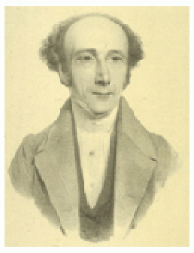 |  |  |
Sir Robert Carswell (1793-1857)
Robert Carswell has long been regarded as the most talented illustrator of pathological conditions to emerge from the early days of taught anatomy in 19th century Britain. His exceptional set of some 1000 watercolours depicting case histories was produced over a period of time, first in the early 1820s as a student and later as a physician and professor. Carswell's talent was spotted early, and under the mentorship of the Edinburgh military surgeon John Thomson he was sent to post-Napoleonic French hospitals to start building a pictorial teaching library of morbid anatomy cases directly from dissected bodies. A few years later as a professor himself at the newly-founded London University, and physician at the University Dispensary (later North London Hospital), he obtained leave of absence to return to Paris and complete his work. Carswell's later career involved a stint as physician to the King of the Belgians, and to King Louis of France in exile in England, for which he was knighted by Queen Victoria.
The Collection
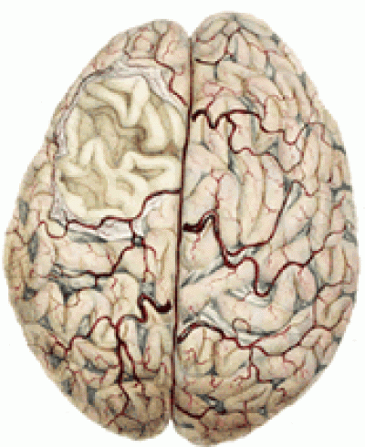
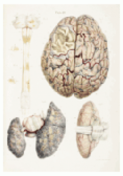
By 1838 Carswell's sketching, painting and collecting activities had culminated in the publication of his major work, Illustrations of the Elementary Forms of Disease, to considerable acclaim, and Carswell had amassed an astonishing 1,025 individual water-colours and pen and ink drawings, or "delineations" as he called them.
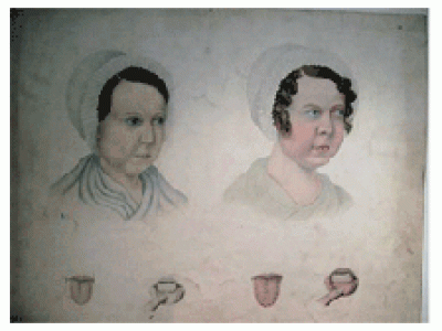
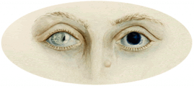
Most of the drawings and watercolours, which range in size from 4 x 4cm to as much as 45 x 30cm and above, are anatomical specimens shown in situ at the post-mortem examinations, illustrating various aspects of the human anatomy, such as an internal organ. Others are of living patients, depicting individual parts such as a limb, or the face. The set is arranged in 17 sections, the largest section (146 drawings) being of the lungs, pleura and bronchi. Many have a description of the pathology, and some the clinical history, written in Carswell's own hand, later copied and bound into volumes.
Research Significance
This unique collection of groundbreaking work is a valuable resource for medical historians and art historians alike as well as students of both disciplines. For the first time ever diseased structures were depicted in colour in detail, allowing medical practitioners as well as teachers and students to learn about the nature of disease, before the advent of microscopy. Pathological anatomy (later pathology) was only just emerging as a medical speciality and contained in the Carswell collection are many items of historical significance, notably the first illustrations of the pathology in Hodgkin's Disease (shown at Hodgkin's classic lecture in 1832), the first portrayal of the lesions on the spinal cord in multiple sclerosis, and the first depictions of iron deficiency anaemia.
The Restoration Project
This important collection has now been conserved and re-housed with a generous grant from the Wellcome Trust. After years of being handled and used in regular teaching, the collection had accumulated significant quantities of dirt and dust, making access difficult and damaging. As the poor condition is an integral aspect of its historical context, it was decided to apply the least invasive and most sympathetic treatment method, so minimum cleaning, deacidification, and repairs were applied. A particular method of inlaying the original sheets was then used to enable viewing form both sides, so that none of the content, either drawn or written, is lost. Re-housed in archival standard solander boxes, the drawings are once again easy to handle and view and made fully accessible for teaching and research.
Descriptive records and digital images of the collection are being created for online use.
This website will be updated and expanded as the project progresses.
 Close
Close

